Case Identification
Procedure: Pinhole® Surgical Technique
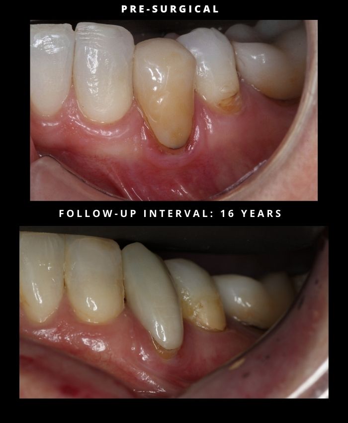
Tooth/Teeth: #20–21
Procedure date: 2010
Post-operative interval: 16 Years
Post-operative examination date: 2026
This case documents a 16-year clinical observation following treatment of localized gingival recession at teeth #20 and #21 using the Pinhole® Surgical Technique.
The pre-surgical photograph was obtained in 2010 and demonstrates localized recession with visible root exposure in the mandibular left premolar region. The follow-up image was captured 16 years post-treatment, allowing evaluation of soft-tissue behavior well beyond early healing, maturation, and intermediate remodeling phases.
At the 16-year post-surgical interval, the treated sites demonstrate:
• Stable coronal gingival margin position relative to the cervical tooth contours
• Maintained soft-tissue thickness and tissue adaptation at the treated teeth
• Favorable gingival contour without visible scarring
• No obvious clinical signs of relapse when comparing available landmarks
Despite differences in camera angle, lighting, and retraction between the two time points, the clinical features permit meaningful comparison of gingival margin position and tissue form. The observed soft-tissue architecture appears stable over an extended interval, suggesting durable tissue behavior at the treated sites.
At this interval, healing and remodeling are considered complete. The gingival margin position and tissue thickness observed reflect long-term biologic stability rather than short-term post-procedural changes.
The clinical significance of this case lies in the documentation of long-term soft-tissue stability maintained over 16 years following a minimally invasive periodontal procedure performed without incisions or sutures.
The Pinhole® Surgical Technique, developed by Dr. John Chao, is a biologically driven approach to the management of gingival recession that emphasizes tissue preservation and long-term stability.
For additional information:
www.pinholesurgicaltechnique.com