Case Identification
Procedure: Pinhole® Surgical Technique
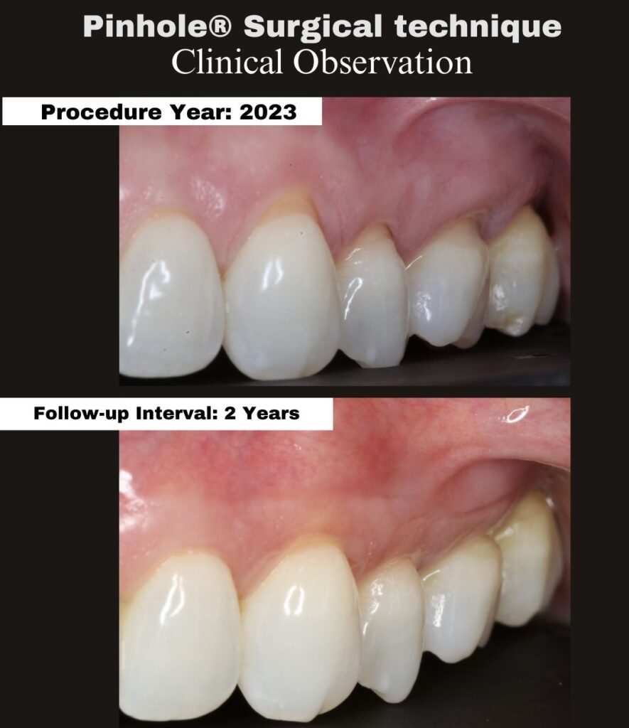
Tooth/Teeth: #2–15
Procedure year: 2023
Follow-up interval: 2 Years
Follow-up examination date: 01/23/26
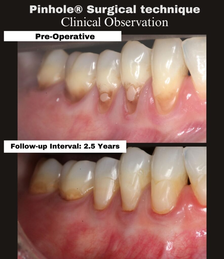
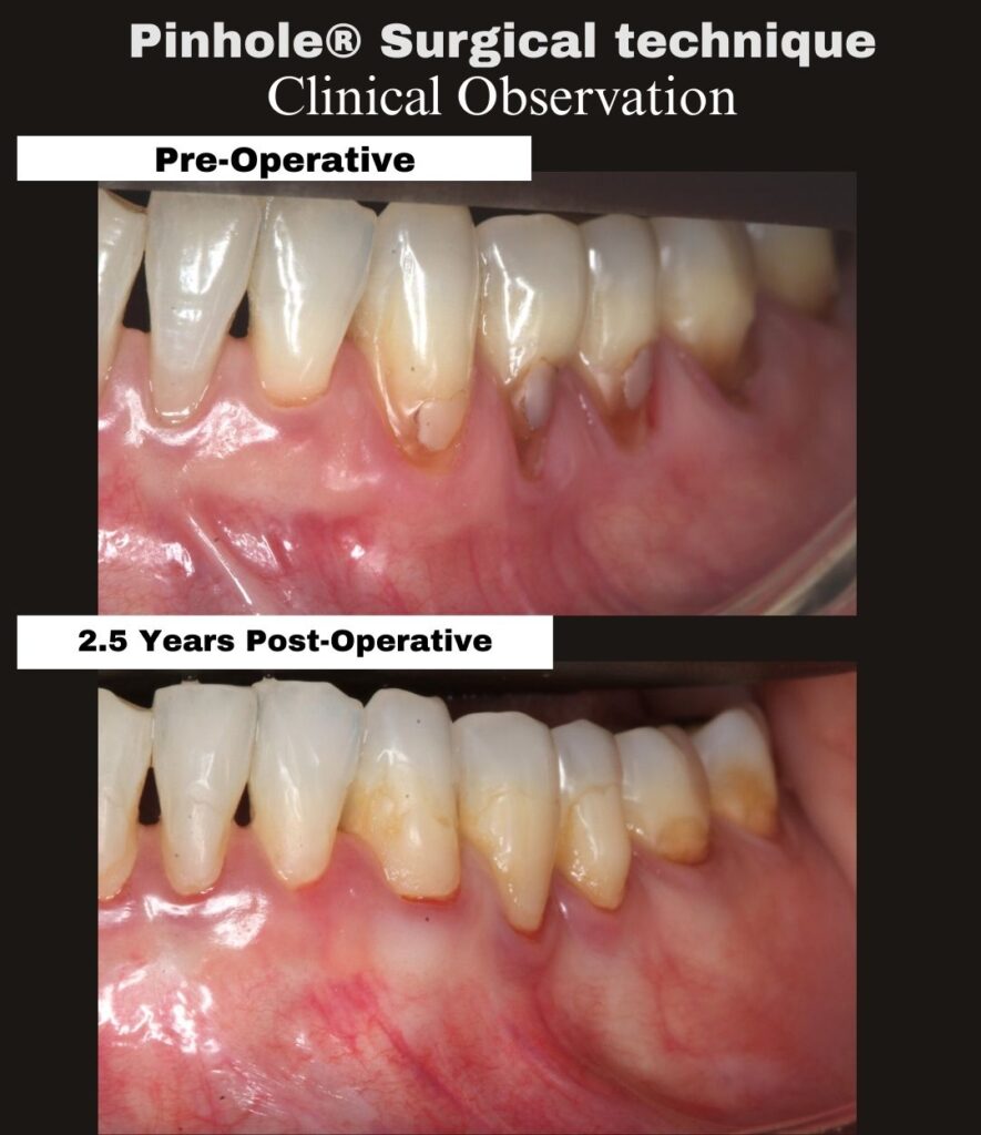
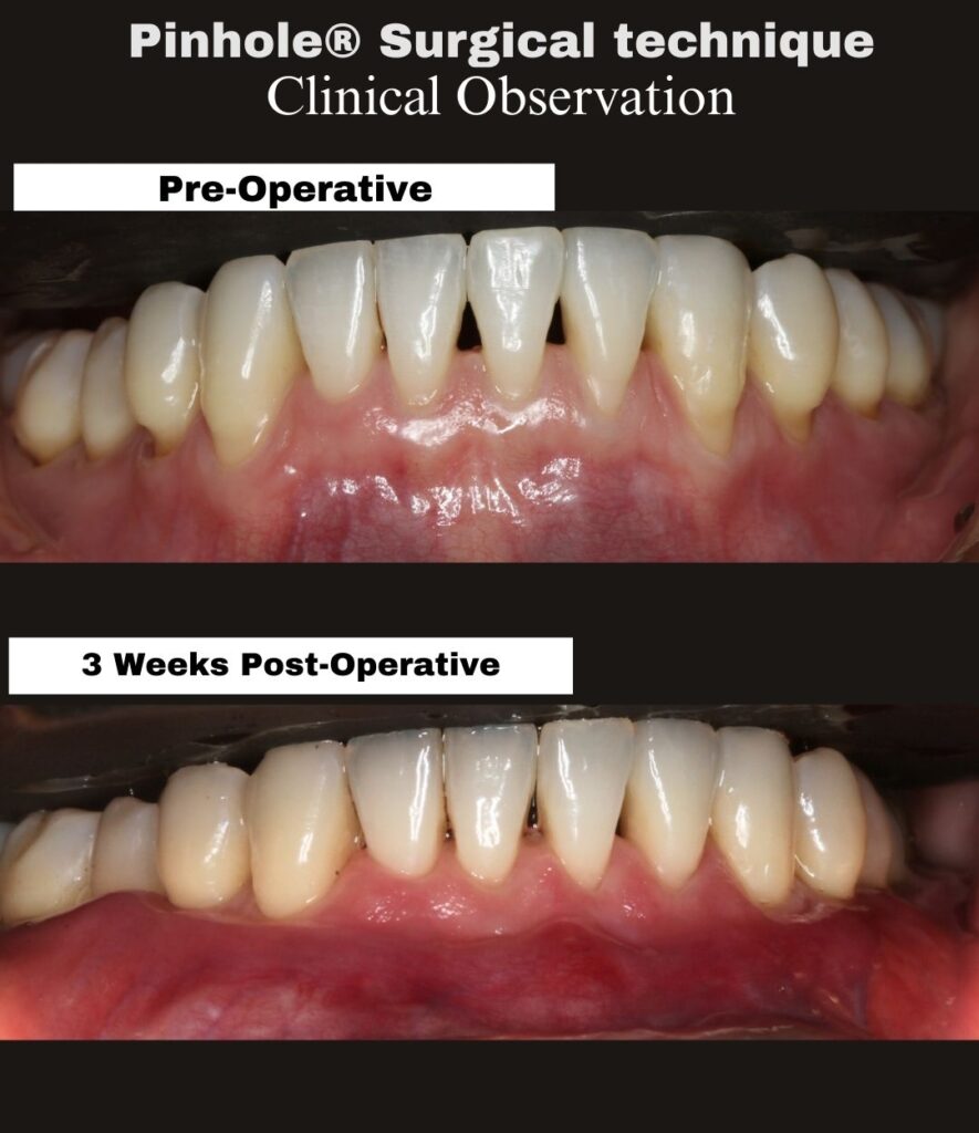
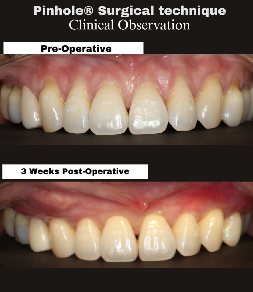
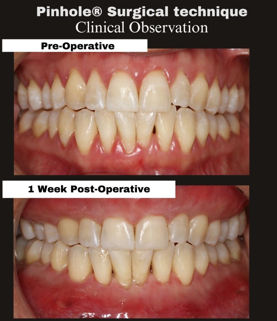
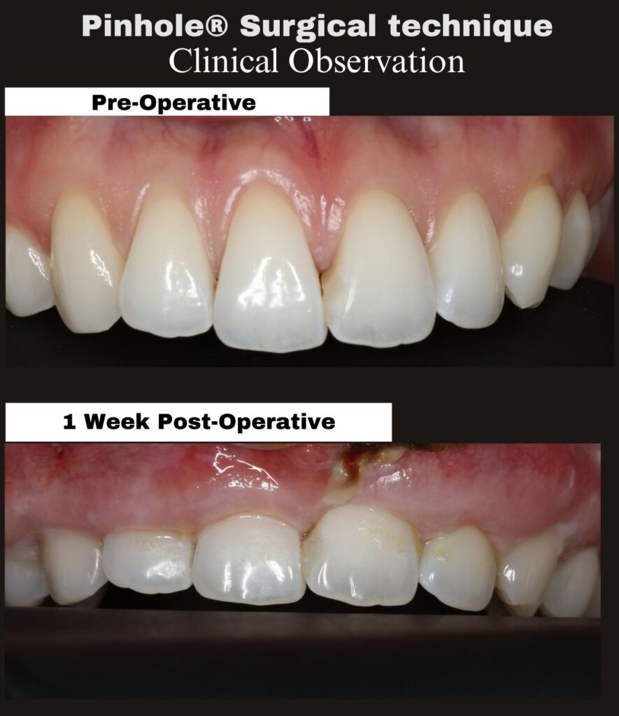
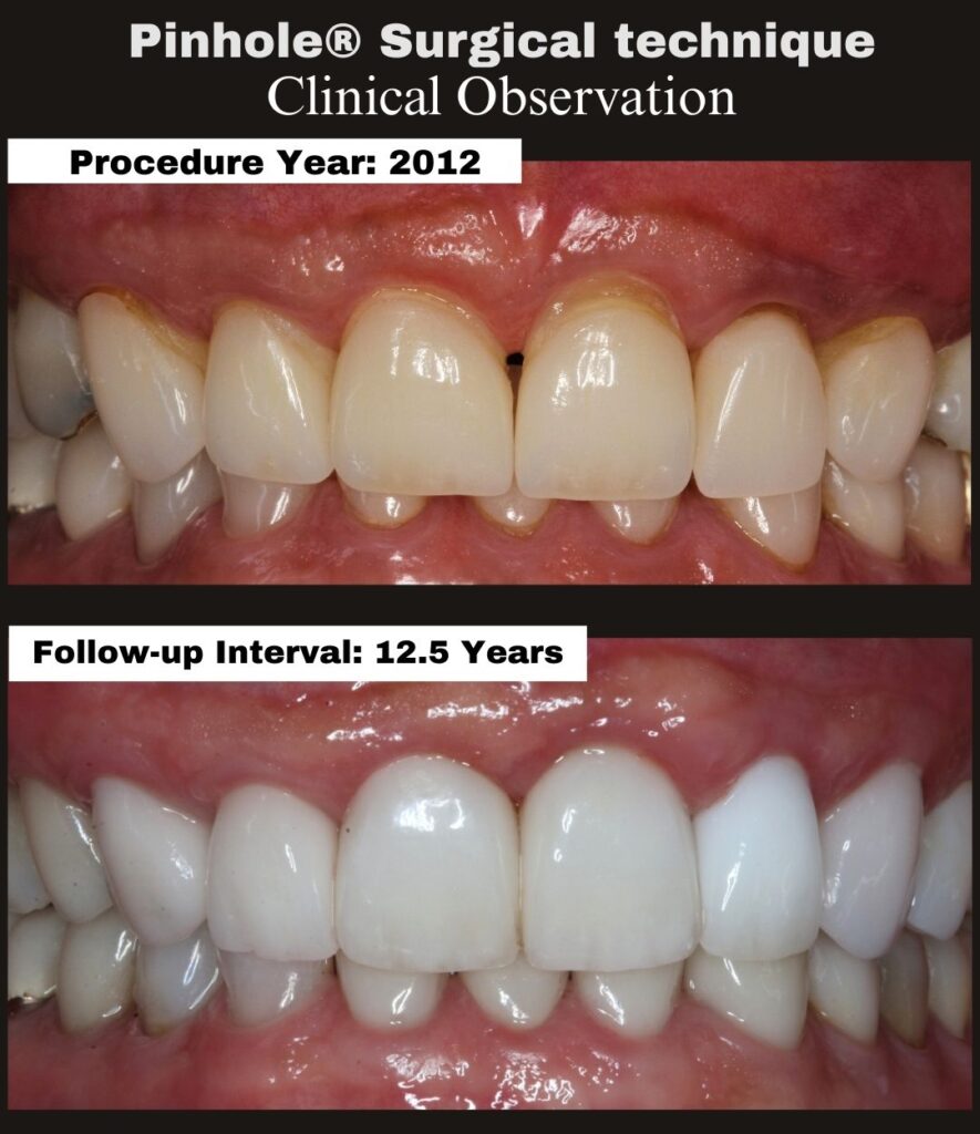
This case documents the 2-year post-operative outcome following treatment of generalized maxillary gingival recession using the Pinhole® Surgical Technique.
As documented in the original operative record from 2023, the primary clinical objective was to achieve adequate soft-tissue release and controlled overcorrection to increase tissue thickness and establish a biologic environment supportive of long-term stability. The gingival margin position at surgery was intentionally not considered the final outcome.
At the 2-year follow-up interval, the treated sites demonstrate:
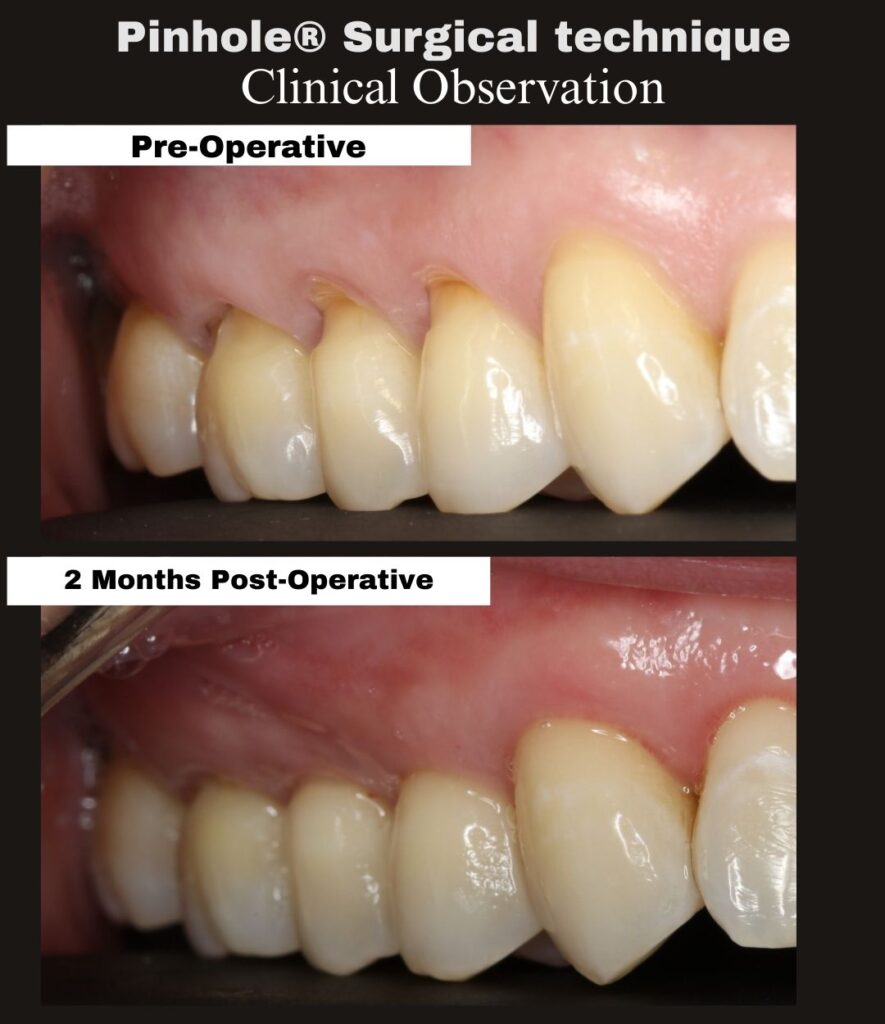
• Sustained coronal gingival margin position
• Stable soft-tissue thickness with maintained volume
• Preserved papillary architecture without scarring
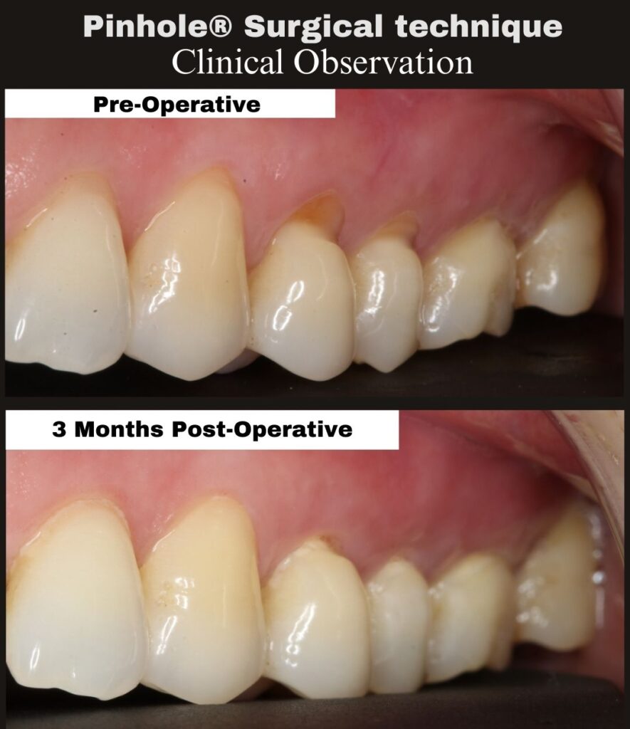
• Long-term tissue adaptation consistent with biologic remodeling
At this interval, tissues have completed maturation and remodeling. The observed gingival margin position and contour reflect the patient’s biologic response over time rather than intraoperative placement. Stability at 2 years supports the principle that controlled overcorrection and adequate tissue release allow the gingiva to settle into a durable, biologically determined position.
The clinical significance of this case lies in the demonstration of long-term stability achieved without incisions or sutures, reinforcing the biologic basis of the Pinhole® Surgical Technique.
The Pinhole® Surgical Technique was developed by Dr. John Chao as a biologically guided, minimally invasive approach to the management of gingival recession.
For additional information:
www.pinholesurgicaltechnique.com
#PinholeSurgicalTechnique #ClinicalObservation #GingivalRecession #LongTermFollowUp #BiologyDrivenHealing #MinimallyInvasivePeriodontics #DrJohnChao