Complete root coverage
77.9%
The percentage of studied Miller Class I–II sites reported with complete root coverage at long-term follow-up.
Published long-term evidence
This retrospective case series followed 28 patients and 68 Miller Class I–II recession sites for an average of 173.8 months.
Study summary
The figures below describe outcomes observed in this study population; they do not predict an individual patient’s result.
Complete root coverage
The percentage of studied Miller Class I–II sites reported with complete root coverage at long-term follow-up.
Mean root-defect coverage
The average percentage of the original recession defect covered at the mean 14.5-year follow-up.
Patient esthetic satisfaction
Patient-reported esthetic satisfaction across the recalled patients at long-term follow-up.
Clinical photographs
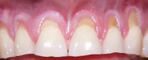
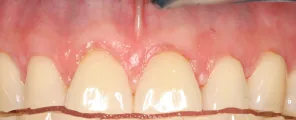
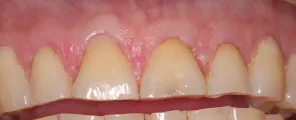
Clinical photographs reproduced from the published case series—the same patient at baseline, 33 months, and the 18-year recall.
Read the source
Use the PubMed record to confirm the study design, inclusion criteria, methods, outcomes, and limitations before citing the findings.
See clinical examples
Review selected multi-year follow-ups, then open the Academy’s complete case gallery.
The complete evidence library
Peer-reviewed research on the Pinhole® Surgical Technique spanning 2012–2026—from the original study and the 14.5-year follow-up to the randomized trial and independent case series from around the world.
Start with the flagships
The original feasibility study, the longest follow-up published to date, and the randomized head-to-head against the grafting gold standard.
The original · IJPRD 2012
Complete root coverage in Class I–II sites, with 94.0% mean defect reduction, a 22.3-minute mean surgery time, and 95.1% patient satisfaction.
Longest follow-up · IJPRD 2025
Mean root-defect coverage maintained at a mean 14.5 years across 68 Class I–II sites—with 77.9% complete coverage.
Randomized trial · Compendium 2025
Split-mouth trial in 36 patients: root coverage and recession reduction statistically equivalent to the connective-tissue graft—without the second surgical site.
By Dr. Chao and collaborators
Original research, the randomized trial, long-term follow-ups, and supporting protocols from the technique’s inventor and his co-investigators.
Int J Periodontics & Restorative Dentistry · 2012 · Chao — the original study: 81.2% complete coverage in Class I–II sites, 94.0% mean defect reduction, 22.3-minute mean surgery time, 95.1% patient satisfaction.
Int J Periodontics & Restorative Dentistry · 2025 · Chao, Reyes Rosales, El Chaar, Shibly, Al-Sabbagh, Ma — 28 patients, 68 Class I–II sites at a mean 14.5 years: 86.6% mean defect coverage, 77.9% complete coverage.
Compendium 46(1) · 2025 · Shibly, Chao, Albandar, Almehmadi, Al-Sabbagh — 36 patients, 1 year: root coverage 63.6% vs 65.4% and recession reduction 1.97 vs 1.98 mm, statistically equivalent.
University at Buffalo, Periodontics · 2020 · Shibly, Chao — the randomized, split-mouth, double-blinded protocol behind the trial: 52 subjects, endpoints through 24 months.
Journal of Cosmetic Dentistry · 2024 · Chao, Shibly, Tso, Ma — PST® combined with surgical crown shortening: complete coverage on maxillary teeth and near-complete on mandibular incisors.
Perio-Implant Advisory · 2025 · Froum, Chao — PRF as a natural scaffold: greater keratinized tissue width, better coverage, and reduced postoperative pain versus PST® alone.
Journal of Clinical Dentistry and Oral Care 4(2) · 2026 · Chao, Mishail, Safa, Reyes Rosales — a custom-fit appliance that protects healing surgical sites while allowing brushing of exposed crowns.
Il Dentista Moderno · 2014 · Chao, Shanelec — the microsurgical origins of PST® and how it compares with free connective-tissue grafting.
Independent research
Investigations by unaffiliated clinicians and universities—including imaging studies, biomaterial comparisons, and multi-site case series.
J Indian Society of Periodontology 21(6) · 2017 · Reddy — independent case series of 18 sites: 96.7% mean root coverage with minimal complications.
Imaging Science in Dentistry 53(1) · 2023 · Shahmirzadi et al. — 3D CBCT imaging after PST® showed an average alveolar bone gain of more than 0.5 mm.
J Esthetic and Restorative Dentistry 36(2) · 2023 · Trivedi, Kolte, Kolte, Deshpande — randomized comparison across 165 sites; adding A-PRF improved depth, width, thickness, and esthetic scores.
Advances in Human Biology · 2024 · Patel et al. — significant 12-month improvement in recession depth, width, and attachment using an amniotic-membrane allograft.
Contemporary Clinical Dentistry 11(1) · 2020 · Agarwal et al. — 20 esthetic-zone sites with T-PRF membrane: 87% root coverage at six months with high patient satisfaction.
Contemporary Clinical Dentistry 10(4) · 2019 · Sharma, Dureja, Arora — two documented cases with complete root coverage and minimal discomfort.
Int J Surgery Case Reports · 2021 · Mostafa, Mandil — propolis root conditioning alongside PST®: enhanced healing and minimal postoperative complications.
Int J Scientific and Research Publications · 2021 · Choudhury et al. — both membranes achieved positive root coverage and gingival regeneration in Class I recession.
Advances in Dentistry & Oral Health 13(1) · 2020 · Mostafa et al. — review of multiple-site treatment in a single appointment without donor sites or graft exposure.
J Dental Sciences and Oral Rehabilitation · 2020 · Gaba et al. — documented case highlighting reduced discomfort and strong esthetic results.
Advances in Dentistry & Oral Health 4(3) · 2017 · Al-Almaie — review reporting up to 94% defect reduction with minimal postoperative complications.
Professional reviews and press
Clinical reviews and professional coverage discussing where PST® fits in practice.
Journal of the California Dental Association · Beck — in-depth professional review of benefits, limitations, patient selection, and long-term considerations.
Dental Economics · 2022 · Froum — decade-long evaluation reporting 81% complete root coverage in Class I–II defects.
A Closer Look · 2018 · Zalesky — how PST® integrates with restorative and esthetic treatment planning.
Colgate · 2023 · medically reviewed consumer overview citing 96.7% root-coverage success and 95% esthetic satisfaction.
For dentists
The free webinar covers the technique, case selection, and implementation—presented by Dr. Chao.