Case Identification
Procedure: Pinhole® Surgical Technique
Tooth/Teeth: #3–14, 18–28, 27–31
Procedure date: 01/23
Post-operative interval: 3 Years
Post-operative examination date: 01/26
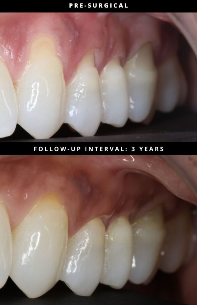
This case documents a 3-year clinical observation following treatment of generalized gingival recession involving both the maxillary and mandibular arches using the Pinhole® Surgical Technique.
The pre-surgical photograph demonstrates generalized recession with visible root exposure and reduced soft-tissue thickness across multiple anterior and posterior teeth. The follow-up image was obtained three years post-treatment, allowing evaluation beyond early healing and tissue maturation and into the long-term remodeling and stability phase.
At the 3-year post-operative interval, the treated sites demonstrate:
• Stable coronal gingival margin position
• Maintained soft-tissue thickness and coverage
• Favorable gingival contour without visible scarring
• Tissue appearance consistent with long-term biologic stability
At this interval, healing and remodeling are considered complete. The gingival margin position and tissue architecture observed reflect sustained biologic response rather than short-term post-procedural effects. The absence of observable relapse over three years suggests durable soft-tissue behavior following treatment.
The clinical significance of this case lies in the documentation of long-term stability across a broad range of teeth following a minimally invasive periodontal procedure performed without incisions or sutures.
The Pinhole® Surgical Technique, developed by Dr. John Chao, is a biologically driven approach to the management of gingival recession that emphasizes tissue preservation, controlled release, and long-term stability.
For additional information:
www.pinholesurgicaltechnique.com