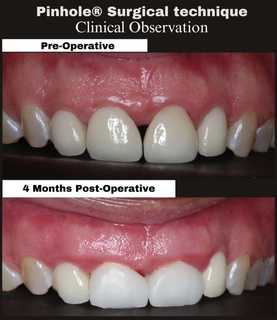
Case Identification
Procedure: Pinhole® Surgical Technique with Papilla Regeneration
Tooth/Teeth: #8–9
Procedure date: 09/02/25
Post-operative interval: 4 Months
Procedure time: 10:51 AM – 12:26 PM (95 minutes)
This case documents mid-term post-operative healing following treatment of maxillary anterior gingival recession with associated interdental papilla deficiency using the Pinhole® Surgical Technique.
As recorded at the time of surgery, the primary clinical objective was to achieve adequate soft-tissue release and controlled overcorrection to increase tissue thickness and create a biologic environment supportive of papilla regeneration and long-term stability. Immediate post-surgical tissue position was not intended to represent the final esthetic outcome.
At 4 months post-procedure, the treated sites demonstrate:
• Re-established interdental papilla between #8 and #9
• Increased soft-tissue thickness and volume in the anterior maxilla
• Stable gingival margins with harmonious tissue contours
• Resolution of post-operative inflammation
• Intact tissue continuity without evidence of incisions or sutures
At this interval, soft-tissue maturation and biologic remodeling are well underway. Gingival margin position and papillary architecture reflect host tissue response over time rather than intraoperative positioning alone, consistent with biologically guided healing.
The clinical focus of this evaluation is assessment of tissue stability, papilla regeneration, and integration of soft tissue within the esthetic zone.
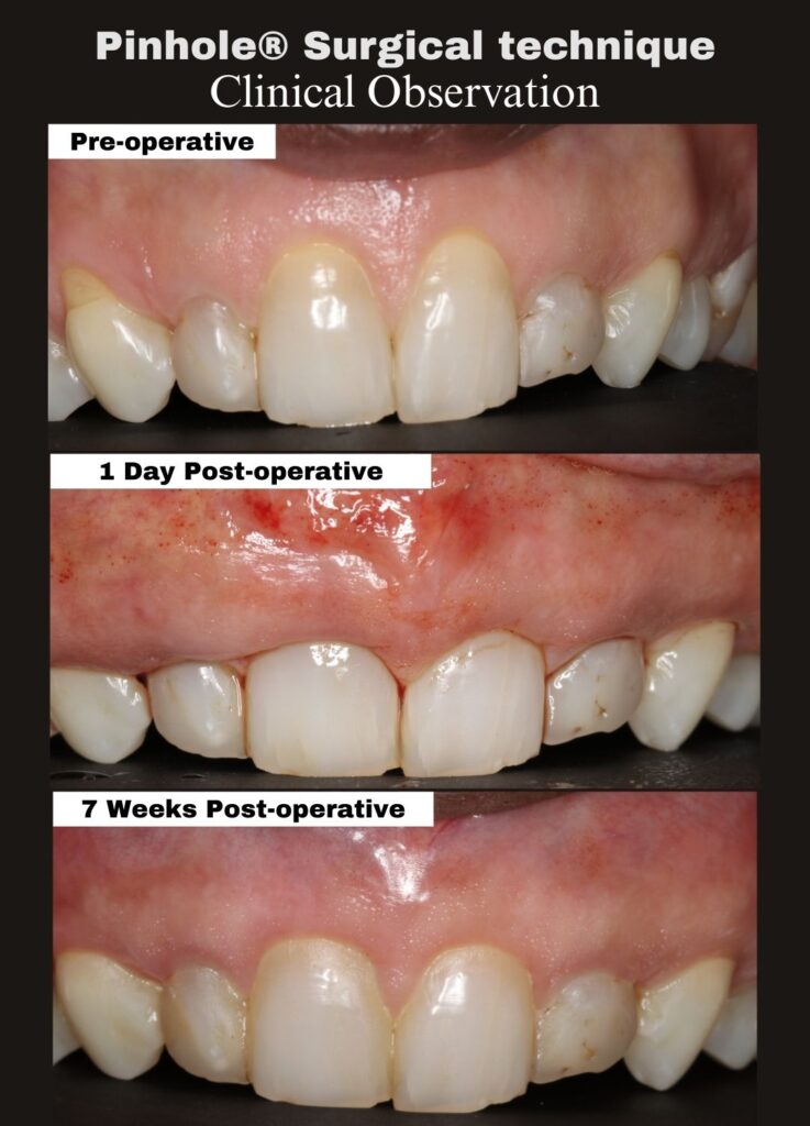
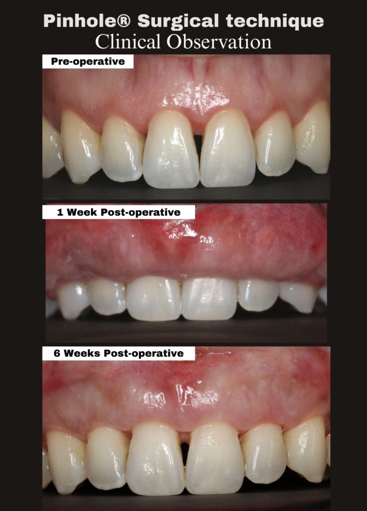
The Pinhole® Surgical Technique is a biologically guided, minimally invasive approach developed by Dr. John Chao for the management of gingival recession and associated soft-tissue deficiencies without scalpels or sutures.
For additional information:
www.pinholesurgicaltechnique.com
#PinholeSurgicalTechnique #ClinicalObservation #PapillaRegeneration #EstheticZone #GingivalRecession #BiologyDrivenHealing #MinimallyInvasivePeriodontics