Case Identification
Procedure: Pinhole® Surgical Technique
Tooth/Teeth: #21–26, 28, 29
Procedure date: 03/28/25
Post-operative interval: 10 Months
Post-operative examination date: 01/28/26
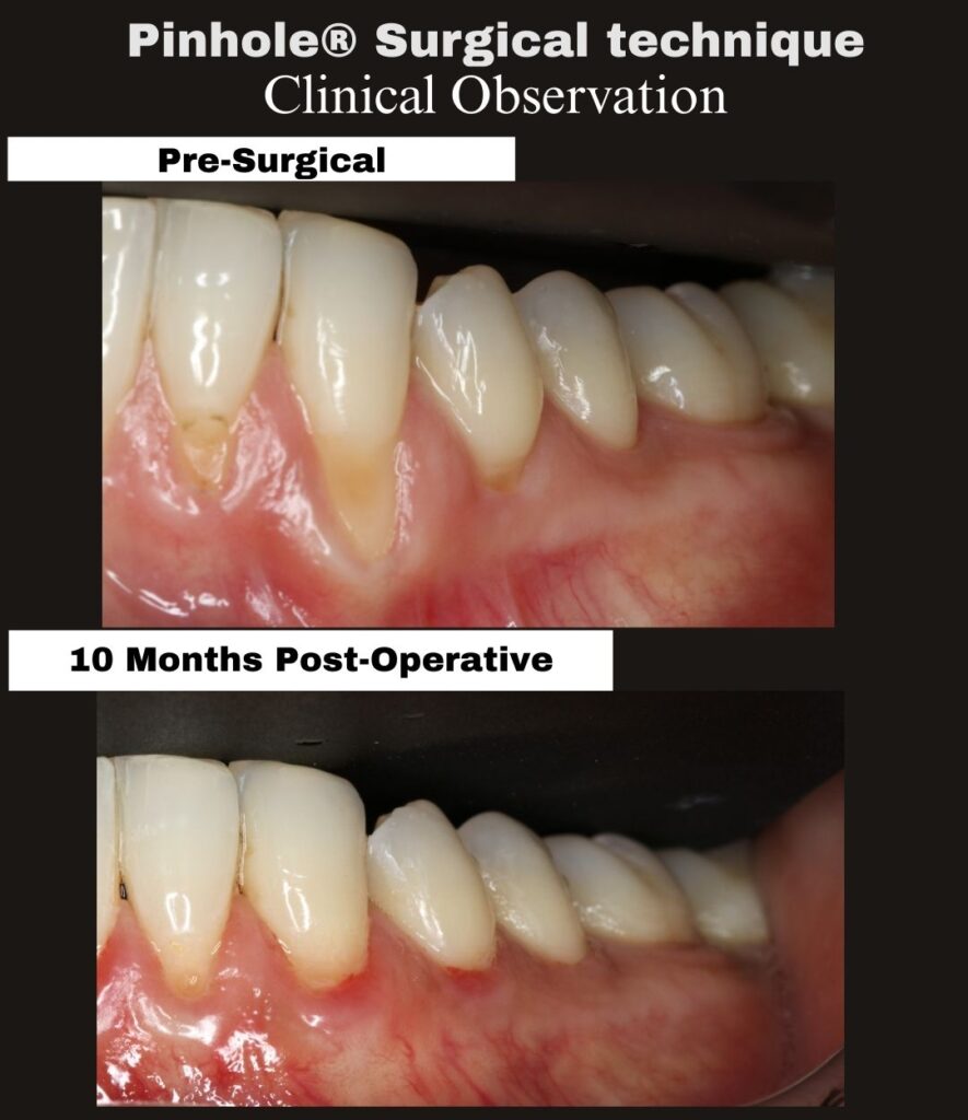
This case documents a 10-month clinical observation following treatment of mandibular gingival recession involving teeth #21–26, 28, and 29 using the Pinhole® Surgical Technique.
The pre-surgical photograph demonstrates gingival recession affecting the mandibular anterior and premolar region, with visible root exposure and reduced soft-tissue thickness. The follow-up image was obtained ten months post-treatment, allowing evaluation beyond early healing and into the intermediate remodeling and maturation phase of soft-tissue healing.
At the 10-month post-operative interval, the treated sites demonstrate:
• Stable coronal gingival margin position
• Increased and maintained soft-tissue thickness
• Favorable gingival contour without visible scarring
• Tissue appearance consistent with ongoing biologic maturation
At this interval, the tissues have progressed beyond early wound healing and are continuing through longer-term remodeling. The observed gingival margin position and tissue architecture reflect biologic stability rather than transient post-surgical changes.
The clinical significance of this case lies in the demonstration of sustained soft-tissue improvement and stability in the mandibular region at an intermediate follow-up interval following a minimally invasive periodontal procedure performed without incisions or sutures.
The Pinhole® Surgical Technique, developed by Dr. John Chao, is a biologically driven approach to the management of gingival recession that emphasizes tissue preservation, controlled release, and long-term stability.
For additional information:
www.pinholesurgicaltechnique.com